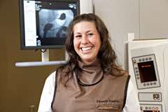
The Diagnostic Imaging Center at Pinnacle Hospital provides a variety of cutting-edge digital imaging technology, including Magnetic Resonance Imaging (MRI), CT Scans (Computerized Tomography), Ultrasound, and Digital X-Ray, together with expert professional interpretation.

The equipment and facilities dedicated to diagnostic imaging at Pinnacle have been designed to provide our patients with the very latest technology in a warm, inviting environment. The staff of the Diagnostic Imaging Center put service and patient comfort above all else as they conduct comprehensive imaging services. By utilizing digital imaging, the results of your imaging session are available immediately for radiology and other specialists to make a proper diagnosis.
The onsite staff of radiologists and radiology technologists offer years of experience in providing a comfortable, convenient experience. Our professionals apply their consummate skills in reading diagnostic results and provide thorough and timely diagnostic images to assist your physician in providing the highest level of care.
Services Available
Diagnostic imaging technology services available at the Diagnostic Imaging Center include: MRI, CT, Digital X-ray, Ultrasound, and Myelography.
MRI (Magnetic Resonance Imaging)

Magnetic resonance imaging (MRI) is a test that uses a magnetic field and pulses of radio wave energy to make pictures of organs and structures inside the body. Because of its advanced technology, MRI images provide physicians with information that cannot be seen utilizing X-ray, ultrasound, or even computed tomography (CT) scan. The images that are produced by an MRI procedure are amazingly clear and allow for the most comprehensive diagnosis of medical abnormalities. An MRI may be prescribed for any number of diagnostic reasons, including scanning the brain and other body parts for tumors, viewing blockages of blood vessels, or scanning bones and related structures for signs of arthritis. Similar to a CT procedure, the patient is placed on a table and the MRI equipment surrounds the patient. Once completed, the images are read by a radiologist with findings relayed to your physician or specialist.
CT (Computed Tomography)
CT or computed tomography uses powerful X-ray technology, often utilizing an iodine dye (contrast material) to create the best 3-D image or the patient’s body. CT provides physicians with the highest level of contrast resolution to create the most vivid images of internal organs. CT procedures are commonly used to detect tumors, blockages in blood vessels, and review actual function of organs by taking a series of images or “slices” that can be viewed in succession, similar to a video. The patient is placed on a table, which is passed through a large ring-shaped device.
Digital X-ray
Traditional X-ray, which passes radiation through the body to project images onto special film, has been updated dramatically with digital X-ray imaging. The images generated through diginal X-ray can be viewed on high-definition monitors by radiologists and physicians to determine diagnosis and treatment options. Because these images are digital, they can be viewed from remote locations or even from around the world.
Myelography
A myelogram procedure utilizes a special dye injected into the spinal canal and X-rays taken to see how the dye fills the space in the spinal canal. This procedure is an excellent method to determine if a patient has disc herniations or pinched spinal nerves. A CT scan often follows after a myelogram to confirm the original diagnosis.
Excellence in Interpretation
The images generated through an MRI, CT, Ultrasound or digital x-ray are often stunningly clear. Yet these images must be reviewed and interpreted by a qualified radiologist to get the very best results.
The Diagnostic Imaging Center at Pinnacle utilizes the services of skilled radiologists to read all images generated by our center. Our radiologists consult directly with physician specialists to develop the best diagnosis for each patient. Pinnacle’s excellence in interpretation is achieved through a multi-disciplinary approach relying on the skills of board-certified radiologists reading the results of images generated by the latest imaging technology.
Having a CT
If you have been told you will be having a CT at Pinnacle Hospital, the following information will provide you with basic information about the procedure, how to prepare, and what to expect.
What is a CT?
CT of computed tomography uses powerful X-ray technology, often utilizing an iodine dye (contrast material) to create the best 3-D image. It provides physicians with the highest level of contrast resolution to create the most vivid images of internal organs. CT procedures are commonly used to detect tumors, blockages in blood vessels, and review actual function of organs by taking a series of images or “slices” that can be viewed in succession, similar to a video. The patient is placed on a table, which is passed through a large ring-shaped device.How to prepare for a CT
Prior to your CT procedure, you will receive general instructions about eating and drinking restrictions. It is advised that you remove all jewelry and accessories before arriving at Pinnacle. Prior to your procedure you will need to remove any hearing aids and removable dental work.Before your procedure, an iodine or contrast agent may be administered. This dye is either administered through a vein or swallowed. Please advise the technician if you have ever had an adverse reaction to an iodine or contrast agent.
Please advise the imaging technician of any of the following:
- You are pregnant
- You are breastfeeding (do not breast feed for two days after the procedure)
- You are allergic to iodine dye
- You have kidney problems - the contrast can damage the kidneys
- You have a history of thyroid problems
- You have diabetes
- You have a heart condition such as heart failure
- You have had a barium enema within four days of the CT
You will remove most articles of clothing, depending on the procedure, and wear a hospital gown during the procedure.
During the CT
You will lie on a table and the CT ring will surround or move around you. It is important that you lie as still as possible to attain the most accurate images. A technician in an adjacent room will conduct the procedure and speak to you throughout the process. Some patients who are uncomfortable in small spaces may be given medication prior to the procedure. The imaging session should last between 30 and 60 minutes. A waiting room is located near the imaging center for family or visitors.After the CT
After your CT, you will get dressed and be able to leave on your own. If you took a sedative before the procedure, an adult will be required to drive you home.Having an MRI
If you have been told you will be having an MRI at Pinnacle Hospital, the following information will provide you with basic information about the procedure, how to prepare, and what to expect.
What is an MRI?
Magnetic resonance imaging (MRI) is a test that uses a magnetic field and pulses of radio wave energy to make pictures of organs and structures inside the body. Because of its advanced technology, MRI images provide physicians with information that cannot be seen utilizing X-ray, ultrasound, or even computed tomography (CT) scan. The images that are produced by an MRI procedure are amazingly clear and allow for the most comprehensive diagnosis of medical abnormalities. An MRI may be prescribed for any number of diagnostic reasons, including scanning the brain and other body parts for tumors, viewing blockages of blood vessels, or scanning bones and related structures for signs of arthritis. Similar to a CT procedure, the patient is placed on a table and the table slides into the MRI machine. Once completed, the images are interpreted by a radiologist with findings relayed to your physician or specialist.
How to prepare for an MRI
Preparing for an MRI test usually requires no special procedures. You will receive general instructions about eating and drinking restrictions. It is advised that you remove all jewelry and accessories before arriving at Pinnacle. Prior to your procedure you will need to remove any hearing aids and dental work.If a contrasting agent is required for your exam, you will be advised. Please advise the technician if you have ever had an adverse reaction to a iodine or a contrast agent.
If any of the following apply to you, you will not be allowed to undergo an MRI:
- You have a pacemaker - an MRI can cause malfunction
- You have shrapnel, bone plates, or pins
- You have aneurysm clips - an MRI may cause the clip to tear the artery it is trying to protect
- You have implanted spinal cord stimulators
- You have inner ear implants
- You have dental implants - some are magnetic
- You have metal heart valves
- You have tattooed eyeliner - iron pigments can cause irritation
- Women who have intrauterine devices (IUD)
- You are pregnant
You will remove most articles of clothing, depending on the procedure, and wear a hospital gown during the procedure.
During the MRI
You will lie on a table that is moved into the MRI machine. It is important that you lie as still as possible to attain the most accurate images. Straps may be used to keep you immobile. A technician in an adjacent room will conduct the procedure and speak to you throughout the process. Some patients who are uncomfortable in small spaces may be prescribed medication prior to the procedure. If you feel this may be an issue with you, please discuss this with your doctor. The imaging session should last between 30 and 60 minutes. A waiting room is located near the imaging center for family or visitors.Having an X-ray
If you have been told you will be having an X-ray at Pinnacle Hospital, the following information will provide you with basic information about the procedure, how to prepare, and what to expect.
What is an X-ray?
Traditional X-ray, which passes radiation through the body to project images onto special film, has been updated dramatically with digital X-ray imaging. The images generated from the process can be viewed on high-definition monitors by radiologists and physicians to determine diagnosis and treatment options. Because these images are digital, they can be view from remote locations or even from around the world.
Preparing for an X-ray
Preparing for an X-ray usually requires no special procedures. You will receive general instructions about eating and drinking restrictions. It is advised that you remove all jewelry and accessories before arriving at Pinnacle.Please advise the radiology technician if you meet any of the following criteria:
- You are pregnant (patients are advised not to have an X-ray while pregnant)
- Have had a barium enema within four days of the X-ray
You may need to remove some articles of clothing, depending on the area of the body that is being X-rayed.
During the X-ray
The radiology technician administering your X-ray will position you to get the optimal image requested by your physician. The X-ray usually only takes a few minutes, but you will be asked to wait while the digital image is generated and reviewed by the technician in case another image is needed to get a better view.


